Decoding Brain MRI Signs of Cryptococcal Meningitis
When cryptococcal meningitis sneaks into the central nervous system, the first clue often comes from a brain MRI. Radiologists don’t just see a blurry picture; they read a story written in contrast enhancement, signal changes, and subtle distortions. Understanding those cues can mean the difference between a missed diagnosis and timely treatment.
Why MRI Is the Modality of Choice
Computed tomography can spot gross hemorrhage or large masses, but it lacks the soft‑tissue resolution needed to tease out the nuances of fungal infection. MRI, with its multiplanar capability and a suite of sequences, highlights the meninges, the ventricles, and the surrounding parenchyma in ways CT simply can’t.
Specifically, T1‑weighted images after gadolinium administration and T2‑fluid‑attenuated inversion recovery (FLAIR) sequences are the workhorses for cryptococcal disease. They show both the direct effect of the organism and the brain’s inflammatory response.
Classic MRI Findings
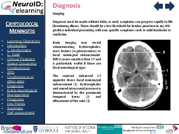
- Leptomeningeal Enhancement: After gadolinium, the pia and arachnoid layers light up in a patchy, sometimes nodular fashion. It’s not the smooth, thick enhancement you’d expect with bacterial meningitis; the pattern is often irregular, reflecting the yeast’s predilection for the subarachnoid space.
- Cryptococcal Gelatinous Pseudocysts: Also called “cryptococcomas,” these are round to oval lesions that appear hyperintense on T2 and FLAIR, but they may not enhance strongly. Their borders can be surprisingly well‑defined, giving the impression of a benign cyst when, in fact, they harbor a stubborn fungus.
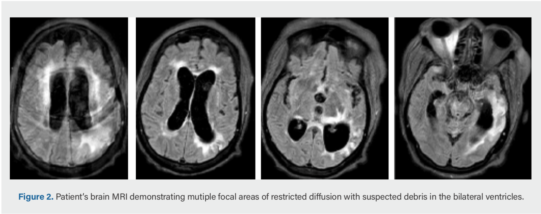
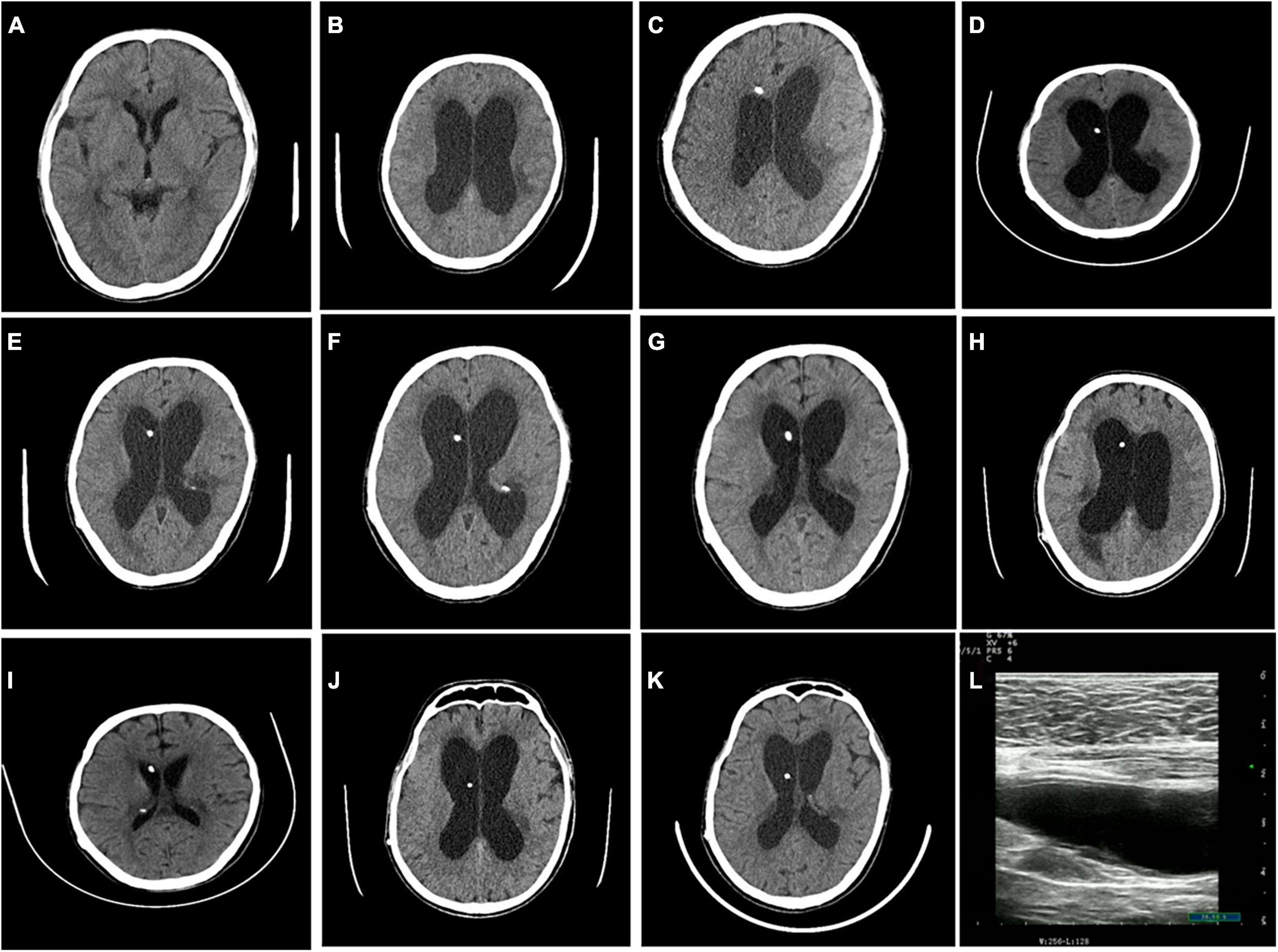
- Ventriculomegaly: Obstructive hydrocephalus is common because the organisms can block CSF flow at the arachnoid granulations or within the foramina. MRI will show enlarged lateral ventricles without a clear mass, a sign that clinicians should not ignore.
- Perivascular (Virchow‑Robin) Space Dilatation: Tiny perivascular spaces may become prominent, especially in the basal ganglia. They appear as tiny, CSF‑like pockets that have taken up the organism, a subtle hallmark that seasoned neuroradiologists recognize.
When the Picture Gets Messier
Not every case follows the textbook. In immunocompromised patients—especially those with advanced HIV—the inflammatory response can be muted. Consequently, enhancement may be faint or even absent, and lesions might look more like ischemic changes.
Conversely, in patients receiving steroids or antiretroviral therapy, a paradoxical inflammatory reaction (sometimes called immune reconstitution inflammatory syndrome) can amplify the enhancement, making the meninges look dramatically inflamed.
Key Sequences and What to Look For
T1‑Weighted Post‑Contrast
Gadolinium highlights disrupted blood‑brain barrier. Look for linear or nodular enhancement along the sulci, especially in the basal cisterns. A “soap‑bubble” appearance—multiple tiny enhancing spots—should raise suspicion.
T2‑Weighted and FLAIR
Both make fluid bright, but FLAIR suppresses the signal from pure CSF, allowing subtle meningeal or periventricular lesions to pop out. Hyperintense lesions in the thalami or cerebellum that don’t follow vascular territories often point to fungal involvement.
Diffusion‑Weighted Imaging (DWI)
While cryptococcal lesions are typically not restricted on DWI, any focal area that does show restricted diffusion warrants a closer look for superimposed bacterial infection or infarction.
Putting Imaging Together With Clinical Context
Radiology never works in a vacuum. A patient presenting with headache, fever, and nuchal rigidity—especially with a CD4 count below 100—needs an MRI interpreted through that lens. If the scan shows basal leptomeningeal enhancement plus ventriculomegaly, the probability of cryptococcal meningitis jumps dramatically.
Conversely, a solitary T2‑hyperintense lesion in an otherwise healthy individual might more likely be a demyelinating plaque. Correlating CSF cryptococcal antigen results with imaging findings clinches the diagnosis.
Management Implications of MRI Findings
Identifying hydrocephalus early can prompt neurosurgical consultation for shunting before irreversible damage sets in. Recognizing cryptococcomas helps guide antifungal therapy duration; larger lesions often require prolonged induction with amphotericin B and flucytosine.
Finally, follow‑up MRIs serve as a barometer for treatment response. A decreasing size of pseudocysts and resolution of enhancement suggest that the fungus is receding, while new or worsening lesions may signal relapse or drug resistance.
Take‑Home Nuggets
- Base‑of‑brain leptomeningeal enhancement is the hallmark—look there first.
- Cryptococcomas are T2‑bright, sometimes non‑enhancing, cyst‑like lesions.
- Hydrocephalus on MRI is a red flag that often demands urgent intervention.
- Immune status dramatically shapes the MRI picture; always ask about HIV or immunosuppressive therapy.
In the end, a well‑read MRI does more than confirm a suspicion; it maps the disease’s territory, predicts complications, and informs the therapeutic roadmap. For anyone grappling with cryptococcal meningitis, mastering those imaging clues can be a lifesaver.