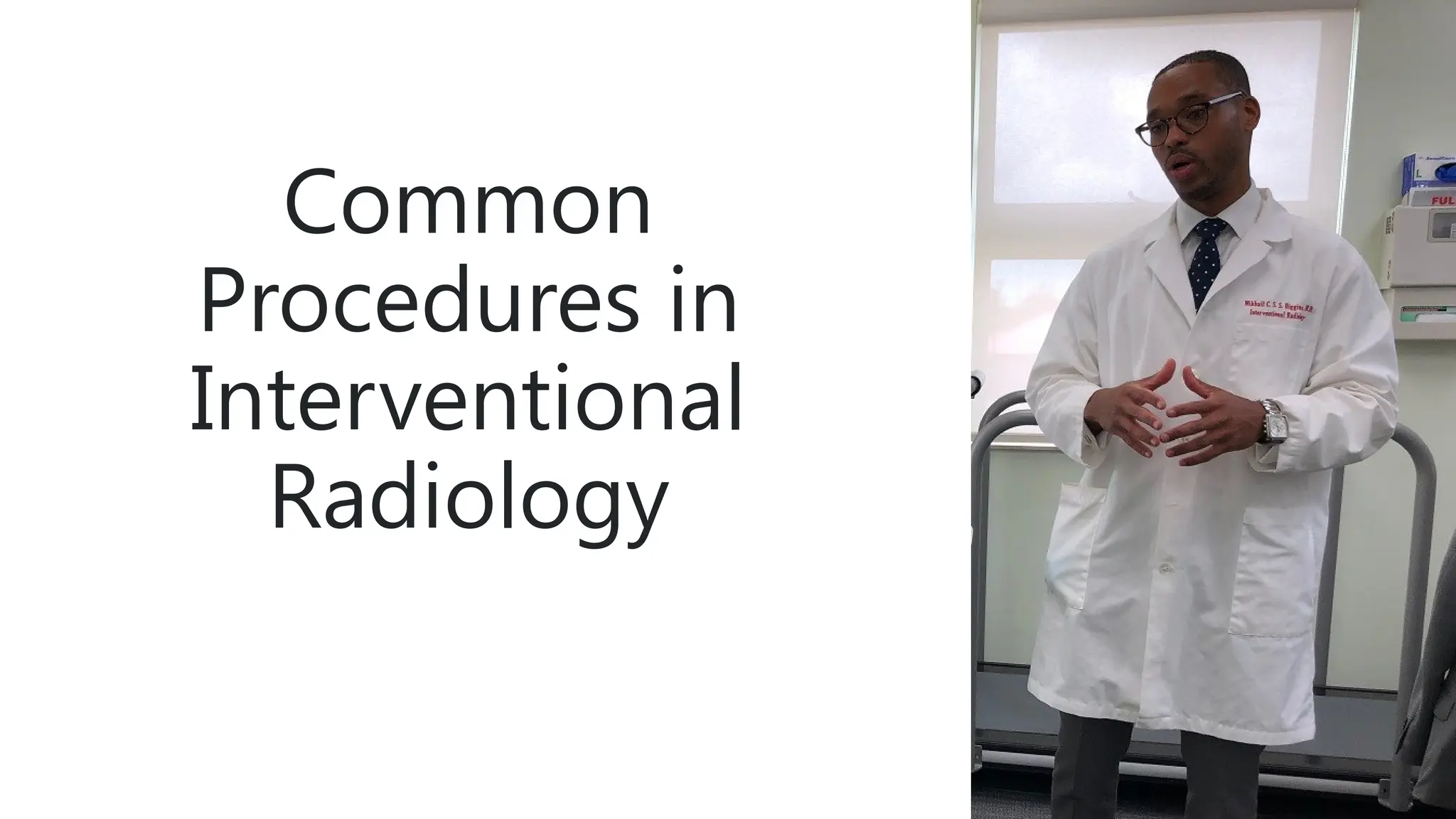
Everything You Need to Know About Radiology Procedures
Why Radiology Is a Cornerstone of Modern Medicine
When a doctor says “let’s get an image,” they’re tapping into a toolbox that can reveal hidden injuries, track disease progress, and guide life‑saving interventions. Radiology isn’t just about pretty pictures; it’s a diagnostic language that translates the body’s internal story into visual data. By visualizing bones, soft tissue, and even blood flow, clinicians can make decisions that would otherwise require invasive surgery.
Beyond diagnosis, many radiologic techniques double as therapeutic platforms—think of fluoroscopy guiding a catheter or interventional radiology stopping a bleeding artery. That dual role makes understanding each procedure essential for anyone navigating the healthcare system.
Core Imaging Modalities at a Glance
- X‑ray: Fast, inexpensive, best for bone and chest imaging.
- Computed Tomography (CT): Cross‑sectional views that excel at trauma, vascular, and oncologic assessment.
- Magnetic Resonance Imaging (MRI): Superior soft‑tissue contrast without ionizing radiation.
- Ultrasound: Real‑time, portable, ideal for obstetrics and superficial structures.
- Fluoroscopy: Continuous X‑ray for guiding procedures in real time.
Each modality has its own sweet spot, and the choice often hinges on the clinical question, patient safety, and resource availability.
CT Scans: Speed Meets Detail
Computed tomography spins an X‑ray source around the patient, capturing multiple slices that a computer stitches into a 3‑D dataset. The result is a rapid, detailed look at complex anatomy—perfect for emergency departments where every second counts.
Typical indications include head trauma, abdominal pain of uncertain origin, and staging of cancer. Modern scanners can complete a chest CT in under a second, delivering images that reveal everything from tiny pulmonary nodules to subtle bone fractures.
Preparing for a CT
- Fast for about 4–6 hours if contrast is used.
- Remove metal objects that could cause artifacts.
- Inform the technologist about allergies or kidney issues.
Contrast agents enhance vascular structures and organ perfusion but carry a small risk of allergic reaction. Radiology teams screen patients carefully to balance diagnostic gain against potential side effects.
MRI: The Soft‑Tissue Specialist
Magnetic resonance imaging uses powerful magnets and radiofrequency pulses to align hydrogen protons, then listens for their return signals. The lack of ionizing radiation makes MRI a go‑to for repeated follow‑ups, especially in younger patients.
Neurology, musculoskeletal, and cardiac imaging benefit most from MRI’s exquisite contrast resolution. For instance, a brain MRI can differentiate between a tumor, inflammation, or demyelinating disease—something a CT might miss.
Key Considerations
- Screen for metallic implants, pacemakers, or cochlear devices.
- Claustrophobic patients may receive mild sedation.
- Gadolinium‑based contrast is generally safe but avoided in severe kidney dysfunction.
Scanning times can stretch to 30–45 minutes, so patients are advised to stay still and follow breath‑holding cues when instructed.
Ultrasound: Real‑Time Imaging at the Bedside
Ultrasound sends high‑frequency sound waves into the body and records the echoes that bounce back. Because it uses no radiation, it’s a favorite for obstetric exams, cardiac function checks, and guiding needle biopsies.
Portable units mean clinicians can perform point‑of‑care scans in the emergency room or intensive care unit, instantly answering questions like “Is there fluid in the abdomen?” or “Is the fetus developing normally?”
When Ultrasound Shines
- Assessing gallbladder stones or liver lesions.
- Evaluating fetal growth and amniotic fluid volume.
- Guiding vascular access or joint injections.
However, ultrasound’s effectiveness depends on the operator’s skill and the patient’s body habitus—obesity can limit image penetration.
X‑Ray and Fluoroscopy: Foundations of Imaging
Traditional X‑ray remains the workhorse for quick assessments—think broken ribs or a chest infection. A single exposure lasts milliseconds, delivering a flat, two‑dimensional view that’s easy to interpret for many common conditions.
Fluoroscopy takes that concept a step further by providing continuous X‑ray imaging, enabling real‑time navigation of catheters, stents, or orthopedic hardware. It’s the visual guide behind procedures such as angiograms and vertebroplasty.
Safety Tips
- Lead aprons protect surrounding tissues during fluoroscopic procedures.
- Minimize exposure time and use low‑dose settings whenever possible.
- Pregnant patients should avoid unnecessary X‑ray unless benefits clearly outweigh risks.
Patient Preparation and Safety Across Modalities
Regardless of the technique, radiology hinges on patient cooperation and safety. Most centers provide written instructions—fasting before contrast, removing jewelry, and disclosing medications. Hydration is often encouraged after contrast studies to help flush the agent from the kidneys.
Radiation dose awareness has grown dramatically. Modern CT scanners incorporate dose‑modulation algorithms that tailor exposure to the patient’s size, while MRI and ultrasound remain radiation‑free alternatives whenever appropriate.
Making Sense of the Images: A Brief Primer
Radiologists act as translators, converting raw data into diagnostic reports. They look for patterns—densities, signal intensities, or flow characteristics—and compare them against normal anatomy. While most reports conclude with a clear impression, they may also suggest further imaging if the picture remains ambiguous.
Patients receiving a report should discuss it with their referring physician, who can contextualize findings within the broader clinical picture. Remember, an image tells a story, but the clinician weaves it into the patient’s unique narrative.
Future Trends Shaping Radiology
Artificial intelligence is already assisting in lesion detection, triaging urgent scans, and even quantifying disease burden. Meanwhile, hybrid machines like PET/CT and PET/MRI merge functional and anatomical data, offering deeper insight into cancer metabolism and brain disorders.
As technology advances, the core principle stays the same: provide clear, actionable information while keeping patients safe. Whether you’re a student, a patient, or a curious reader, understanding the basics of radiology empowers you to ask the right questions and make informed health decisions.