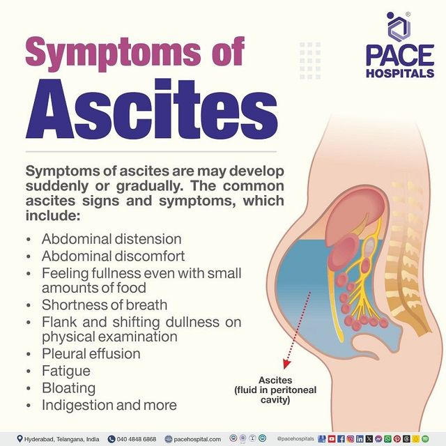
How to Detect and Diagnose Ascites: A Practical Guide
What Is Ascites and Why It Matters
Ascites is the abnormal buildup of fluid in the peritoneal cavity, most often a sign of advanced liver disease but also possible with heart failure, cancer, or infections. While the swelling can be subtle at first, it frequently signals that underlying pathology is progressing and needs prompt attention. Early detection gives clinicians a better chance to address the root cause before complications like spontaneous bacterial peritonitis arise.
Key Clinical Clues in the Physical Exam
During a routine exam, a distended abdomen that feels “full” or “tight” should raise suspicion. Physicians listen for a dullness to percussion that shifts when the patient changes position—a classic sign of fluid moving within the abdomen. Additionally, a “fluid wave” test—where a tap on one side creates a ripple felt on the opposite side—can help confirm the presence of free fluid.
Simple Bedside Tests You Can Perform
- Shifting dullness: Patient lies supine, the examiner percusses from the abdomen’s edge inward; as the patient rolls onto the opposite side, the area of dullness shifts.
- Fluid wave: With both hands placed on the abdomen, a gentle tap on one flank produces a wave that can be felt on the other side.
- Abdominal girth measurement: Comparing the circumference at the level of the umbilicus over time can document progression.
Laboratory and Imaging Tools
When physical findings suggest ascites, imaging usually follows. An abdominal ultrasound is the first‑line modality—quick, non‑invasive, and highly sensitive for even small fluid volumes. It can also reveal liver texture, portal vein diameter, and any masses that might be contributing. If the ultrasound is equivocal or if a more detailed map is needed, a contrast‑enhanced CT scan provides three‑dimensional detail but involves radiation and contrast exposure.
Blood work complements imaging. Liver function tests, serum albumin, and renal panels help gauge the severity of underlying disease. A complete blood count can hint at infection, while coagulation studies are essential before any invasive procedure.
Interpreting Paracentesis Results
The definitive step in diagnosing the cause of ascites is a diagnostic paracentesis—removing a small sample of fluid with a needle under sterile conditions. The fluid is then analyzed for cell count, protein concentration, and albumin level. The serum‑ascites albumin gradient (SAAG) is particularly informative: a SAAG ≥ 1.1 g/dL points toward portal hypertension (most often cirrhosis), whereas a lower SAAG suggests infections, malignancy, or pancreatic disease.
Additional tests may include cytology for malignant cells, cultures for bacterial growth, and triglyceride measurement if a chylous (milky) fluid is suspected. The results guide treatment—antibiotics for infection, diuretics for portal‑hypertensive fluid, or oncologic referral for malignant ascites.
When to Seek Specialist Care
If the fluid is large, recurrent, or accompanied by pain, fever, or altered mental status, a gastroenterologist or hepatologist should be consulted promptly. Patients with low blood pressure, severe renal impairment, or coagulopathy need hospital admission before paracentesis to avoid complications. Moreover, refractory ascites—fluid that does not respond to standard diuretic therapy—often warrants evaluation for transjugular intrahepatic portosystemic shunt (TIPS) or even liver transplantation.
Frequently Asked Questions
- Can ascites develop without liver disease? Yes. While cirrhosis accounts for most cases, heart failure, peritoneal carcinomatosis, tuberculous peritonitis, and pancreatic disorders can also cause fluid accumulation.
- Is an ultrasound always necessary? Not strictly. In a clear clinical picture with obvious abdominal distention, a bedside fluid wave may suffice initially, but imaging is strongly recommended to rule out other intra‑abdominal pathologies and to guide safe paracentesis.
- How often should fluid be re‑checked? For patients on diuretics, a weekly abdominal girth measurement or repeat ultrasound every 1–2 months helps monitor response. Sudden weight gain of more than 2 kg in a few days warrants re‑evaluation.
- Are there non‑invasive alternatives to paracentesis? Emerging techniques like elastography can estimate portal pressure, but they cannot replace fluid analysis for definitive diagnosis. Paracentesis remains the gold standard for determining the underlying cause.