How a CT Scan Works: Everything You Need to Know
What Exactly Is a CT Scan?
A computed tomography (CT) scan is a medical imaging technique that creates detailed cross‑sectional pictures of the body. Unlike a regular X‑ray, which flattens everything onto a single plane, a CT scanner rotates around you, capturing dozens of slices that a computer stitches together into a 3‑D view.
Doctors rely on this depth of detail to spot problems that might be hidden in bone, soft tissue, or blood vessels.
The Science Behind the Images
Inside the machine, an X‑ray tube fires a narrow beam while detectors opposite it measure how much radiation passes through. Different tissues absorb X‑rays to varying degrees—bone blocks more than muscle, and blood‑filled spaces sit somewhere in between. By measuring those differences across dozens of angles, the computer calculates attenuation values and translates them into grayscale pixels.
The result is a stack of “slices” that can be viewed individually or stacked to form a 3‑D reconstruction. Radiologists often scroll through these slices, looking for anomalies that might be invisible on a plain X‑ray.
When Might Your Doctor Recommend a CT?
- Trauma assessment: After a car accident or a fall, a CT can quickly reveal internal bleeding or fractures.
- Cancer detection and monitoring: Tumors show up as distinct densities; CT helps stage disease and watch treatment response.
- Chest and lung evaluation: It can pinpoint infections, pulmonary emboli, or chronic conditions like emphysema.
- Abdominal investigations: From kidney stones to appendicitis, CT offers a fast, comprehensive look.
- Guidance for procedures: Interventional radiologists use real‑time CT images to place needles or drains.
Preparing for Your Scan
Most CT exams require little preparation, but there are a few things to keep in mind:
- Contrast material: Some scans need an iodine‑based dye injected, swallowed, or administered rectally to highlight blood vessels and organs.
- Fasting: If contrast is used, you may be asked not to eat or drink for a few hours.
- Clothing and accessories: Wear loose, metal‑free clothing. Remove jewelry, watches, and belts that could distort the image.
- Pregnancy: Let the technologist know if you’re pregnant or might be; they’ll weigh the benefits against the very low radiation risk.
What Happens During the Procedure?
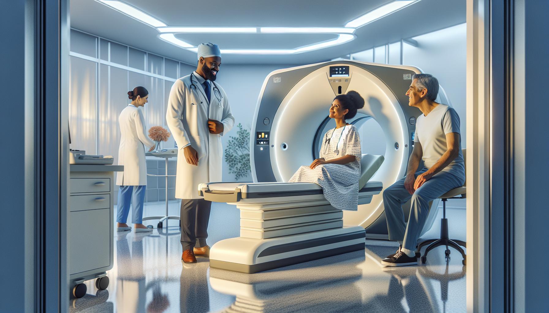
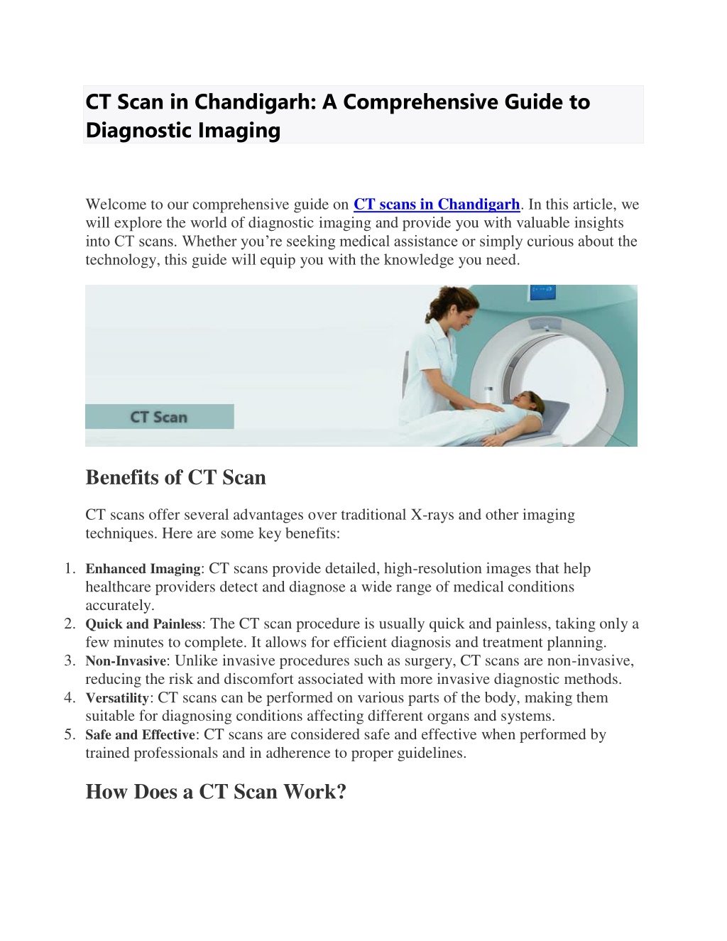
First, you’ll lie on a motorized table that slides into the doughnut‑shaped gantry. If contrast is required, a nurse places an IV line in your arm. The technologist steps out of the room, but stays in contact via an intercom.
When the scan starts, you’ll hear a gentle whir as the X‑ray tube rotates. You’ll be asked to hold your breath for short bursts—usually just a few seconds—to avoid motion blur. The entire exam typically lasts between 5 and 30 minutes, depending on the area being scanned and whether contrast is used.
Understanding Radiation Exposure
CT does involve ionizing radiation, which can theoretically increase cancer risk if overused. However, modern scanners employ dose‑reduction technologies such as automatic exposure control and iterative reconstruction, which keep the dose as low as reasonably achievable.
For perspective, a single abdominal CT might deliver roughly the same amount of radiation as a few years of natural background exposure. Your doctor will only order a CT when the diagnostic benefit outweighs the small risk.
Interpreting the Results
After the scan, a radiologist examines the images and writes a report. This can take anywhere from a few hours to a couple of days. The report will describe any findings—such as masses, fractures, or inflammation—and often suggest next steps, whether that’s further imaging, a biopsy, or a referral to a specialist.
Never hesitate to ask your referring physician to explain the terminology; phrases like “hyperdense lesion” or “ground‑glass opacity” can sound intimidating, but they’re just precise ways of describing what the scanner sees.
Common Myths Debunked
- Myth: “CT scans are always dangerous.”
Fact: The risk is low, especially with modern dose‑saving features. - Myth: “You’ll feel a lot of pain from the contrast.”
Fact: Most people experience a brief warm sensation; severe reactions are rare. - Myth: “The images are blurry if you move.”
Fact: Technologists give clear breath‑hold instructions, and the scanner can compensate for minor motion.
Tips for a Smooth Experience
- Arrive a little early to complete paperwork and discuss any allergies.
- Tell the staff about any metal implants, pacemakers, or recent surgeries.
- If you’re nervous, ask if you can listen to music through headphones during the scan.
- Stay hydrated after a contrast study to help your kidneys flush the dye.
Future Directions: What’s Next for CT Technology?
Researchers are pushing the envelope with ultra‑low‑dose scanners that rival traditional X‑ray quality while preserving 3‑D detail. Dual‑energy CT can differentiate materials—like distinguishing gout crystals from calcium—without extra contrast. Artificial intelligence is also stepping in, helping radiologists flag subtle abnormalities faster.
These advances promise sharper images, less radiation, and quicker diagnoses—meaning patients will benefit from even more precise, patient‑friendly scans.